- MITR Hospital
- 0 Comments
How is Antenatal Hydronephrosis Diagnosed and Monitored?
Antenatal Hydronephrosis is a condition where a fetus develops kidney swelling due to urine buildup. It is commonly diagnosed via prenatal ultrasound. While some cases resolve on their own, others require close monitoring and intervention. Understanding how Antenatal Hydronephrosis diagnosed and monitored is essential for parents and healthcare providers.
Table of Contents
What is Antenatal Hydronephrosis?
Antenatal hydronephrosis refers to the dilatation of the renal pelvis in the fetus, which can be detected through routine ultrasound scans. This condition occurs when outflow of urine from the kidneys to the ureter is obstructed or slowed down.
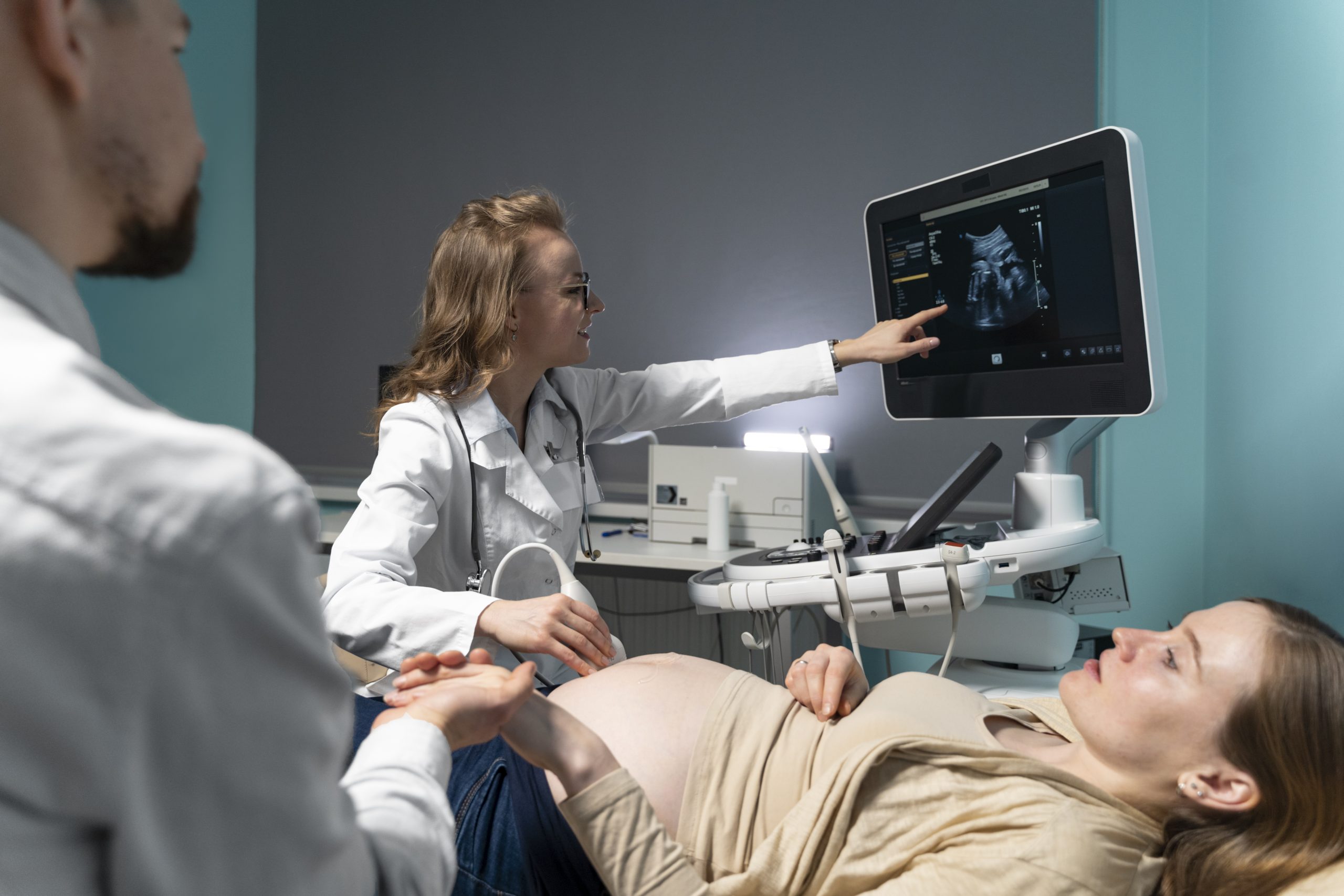
Investigations done for Antenatal Hydronephrosis
1. Prenatal Ultrasound : The primary method for diagnosing antenatal hydronephrosis is through a prenatal ultrasound performed during pregnancy. This imaging test helps detect any swelling in the fetal kidneys and measure the degree of dilation and monitor the swelling throughout the pregnancy.
2. Amniotic Fluid Assessment :
Ultrasound can also tell us about the amniotic fluid levels since reduced amniotic fluid along with hydronephrosis can indicate severe urinary tract obstruction. Proper fluid levels are essential for fetal kidney function and lung development.
3. Postnatal Ultrasound :
Once the baby is born, a follow-up postnatal ultrasound is performed between 7-10 days of life. This helps confirm whether the hydronephrosis persists or has resolved. In cases of bilateral severe hydronephrosis, early ultrasound within the first 48 hours of life is advised.
4. Voiding Cystourethrogram (VCUG) :
If postnatal ultrasound shows persistent hydronephrosis along with dilatation of the ureter or the child has recurrent urinary tract infection, a VCUG test may be conducted to check for vesicoureteral reflux (VUR), a condition where urine flows backward from the bladder to the kidneys.
5. Nuclear Renal Scan (DMSA or EC Scan) :
A renal scan helps assess kidney function, detect any blockages in the urinary tract and assess any scars in the kidneys. EC scan is done to rule out PUJ obstruction or VUJ obstruction and DMSA is done in cases of VUR(vesicoureteric reflux) to check the scars in the kidney.
When to Seek Medical Attention?
- Recurrent urinary tract infections (UTIs)
- Poor urine stream associated with dribbling of urine
- Straining while passing urine
- Palpable kidney which is felt as swelling in the abdomen
FAQs How is Antenatal Hydronephrosis Diagnosed and Monitored?
Antenatal hydronephrosis is a condition in which a fetus develops swelling in the kidneys due to the buildup of urine.
It is usually detected during routine prenatal ultrasound scans performed during pregnancy.
The condition occurs when the flow of urine from the kidneys to the ureter is blocked or slowed down.
Prenatal ultrasound helps doctors identify kidney swelling, measure the degree of dilation, and monitor the condition throughout pregnancy.
Amniotic fluid assessment helps evaluate fetal kidney function, and low fluid levels may indicate severe urinary tract obstruction.
A postnatal ultrasound is done after delivery to check whether the hydronephrosis has resolved or is still present.
A Voiding Cystourethrogram (VCUG) is a test used to detect vesicoureteral reflux (VUR), where urine flows backward from the bladder to the kidneys.
A nuclear renal scan helps evaluate kidney function, detect urinary tract blockages, and identify kidney scarring.
Parents should consult a pediatric urologist or nephrologist if the child develops recurrent UTIs, poor urine flow, straining during urination, or abdominal swelling.
Yes, some mild cases may resolve naturally, while others may require monitoring and medical treatment after birth.